Pressure Point Offloading in the Diabetic Foot

Laurence Foley

Summary

The formation of diabetic wounds has been discussed, with an emphasis on the effects of force on the plantar tissues and the theoretical response of those tissues. As pressure reduction is an integral part of the healing process for diabetic wounds, many types of offloading strategies in the medical literature have been examined. All report success – to varying degrees – but not all comparisons have been applied in a similar way and few have controlled for similar variables. This article presents for discussion a range of off-loading methods that wound care practitioners can employ, depending on the site and severity of the presenting wound.

Introduction

Foot wounds are one of the most common and significant complications associated with chronic diabetes. It has been estimated that 5 per cent of those with diabetes will experience a foot ulcer; indeed, 1.5 per cent of diabetic individuals have a foot wound at any point in time 1. Such wounds can be classed as neuropathic, vasculo-neuropathic or vascular in origin. Since neuropathy is estimated to be present in one in four diabetics 1 and in more than 80 per cent of diabetic patients with foot wounds 2, the majority of diabetic wounds will thus be neuro- pathic in nature and occur on the plantar aspect of the foot 3- 7. Estimates of the prevalence of plantar wounds range from 25-35 per cent of the first toe through 23-50 per cent of the first metatarso-phalangeal joint to 38 per cent for the lesser metatar- sal heads 3-6. Neuropathy produces changes that include sensory, motor and autonomic components 8, all of which will impact on the diabetic foot. “A key factor in the diabetic foot is that dy-namic pressures are higher than in those without diabetes” 9, 10. The causes of increases in dynamic pressures are thought to in-clude bony deformity 11, retraction of the toes, pes cavus 12, lack of soft tissue cushioning, callus formation 13, 14 and limited joint mob- ility 15. The loss of protective sensation (LOPS) from peripheral neuropathy leads to silent injury from biomechanical stresses

Laurence Foley DipCh MSc FAPodA

Laurence Foley Podiatry Department

Fremantle Hospital

Alma Street, Fremantle WA 6160

Telephone: (08) 9431 2342

Facsimile: (08) 9431 2918

when higher pressures are present. Effective pressure reduction strategies are essential in healing and preventing foot wounds.

Forces Acting on the Foot

On the plantar aspect of the foot, the soft tissue between the epidermis and bone assists as part of the cushioning process, protecting the body from the severe mechanical stress exper- ienced by the skin 16. In the diabetic foot, with fat-pad atrophy, digital retraction of the toes, LOPS and autonomic changes leading to a reduction in skin tone, these stresses tend to be magnified. In the biomechanics of biological structures, force is independent of the area over which it is applied and pressure is the applied force divided by its area of application. Thus, the same magnitude of force under one foot can result in different forefoot pressures, depending on the area of application. Birke and Sims 17 refer to four variables that interact to produce com- bined loading under the foot: magnitude, duration, direction (of the forces) and the area over which they load.

Magnitude

While the aspect of diabetics having higher foot pressures than non-diabetics has been examined elsewhere 5, 7, 11, 16, 18-22, the minimum pressure threshold thought to cause ulceration is in dispute – estimates range from 40 to >98 N/cm2 3, 10, 23, with some pressures measured as >110 N/cm2 for ulcers already present 10.

Area

The pressure generated through the foot is a function of the force maintained through a defined area, with the pressure dir- ectly proportional to the force and inversely proportional to the area 11. Therefore, as peak pressures are generated in the foot

102 Primary Intention

August 1999

during forefoot loading and propulsion, high pressure is deliver- ed through a small, rapidly decreasing contact area. If force is reduced at a bony site with an off-loading strategy it must be directed elsewhere, to suitable under-used areas of the foot; that is, to a larger surface area 23.

Duration

Although studies have shown that a relationship exists between moderate repetitive stress and plantar wounds, “ ... there is no consensus on the load duration required to cause ulcerations

... no studies have demonstrated a direct mechanism whereby ulcerations are produced in response to a specific, characteristic mechanical load” 24. Landsman et al 24 suggest that diabetic tissues may be more susceptible to mechanical injury and more sensitive to the rate of deformation than the magnitude of the load itself.

Direction

The direction of the forces in tissues is important, since tissue strength is not the same for all loading patterns. While five loading patterns – tension, compression, bending, shear and torsion – are described for all materials, three have been refer- red to more often in the diabetic literature 10, 25. They are described as surface forces: normal (tension and compression) and shear. Normal forces are applied perpendicularly to a body to cause tension or compression, whereas shear forces cause sliding between parallel planes. When applied, these forces can cause a deformation response referred to as strain; like surface forces, there are three types of strain: tensile, compressive and shear.

There is an internal reaction to these external forces, to maintain equilibrium and resist deformation. It is referred to as stress; that is, the force per unit area acting in a given plane within a material (expressed in N/m2 or Kg/cm2). There are also three types of stress: tensile, compressive and shear.

Normal stresses resist either compression or tension per- pendicularly within a body, while shear stresses resist sliding between parallel planes in a body. Stress is the internal force that develops to resist the strain produced by an externally applied force. If the external force overcomes the internal stress, tissue deformation occurs and, if left unchecked, will cause a wound to develop. (Note: normal and shear stress always exist in com- bination, according to the state of the loading 26.)

In the presence of the four variables – magnitude, duration and direction of the forces acting on the same area – there are

likely to be three types of injury mechanisms in the insensitive foot:

pressure-induced ischaemia;

overt trauma, and

repetitive stress 16.

Concepts of Off-loading

Mechanical protection of the foot is essential for healing 26; con- sideration must be paid to either ‘unweighting’ the foot (that is, no weight on the foot or wound) or ‘off-loading’ (that is, rebalancing the weight on the foot/leg, with the patient still weight-bearing). Guzman et al 27 outline a number of ‘ideal’ characteristics of successful pressure-relieving strategies. They:

provide effective pressure reduction from the ulcer at all times;

have wide application to all patients;

cause no side-effects or secondary lesions;

are easily applied;

encourage patient compliance;

are cost-effective, and

allow other treatment goals to be pursued.

Off-loading Strategies

Callus debridement

The formation of callus, a reactive mechanism of the tissues to shearing and normal strain on the skin, is one of the main precursors to ulcer formation 9. Callus alone has been shown to increase local pressure by up to 30 per cent 28, so effective callus prevention and debridement are vital in reducing the formation of subcutaneous haemorrhaging and ulcer formation. A study of diabetics found there was a relative risk 11 of developing an ulcer under a callused area compared to a non-callused area and the association was much greater than for increased plantar pressures alone 28.

Accommodative padding

Various materials have been used to temporarily off-load pressure on the foot. They can be applied around acute or chronic wounds, depending on the size, site, type and status of the

103 Primary Intention

August 1999

wound. However, care must be taken not to eliminate pressure in one area merely to overload another 29. Armstrong, Liswood and Todd 30 examined accommodative padding cut with an aper-ture to fit around an area on the plantar surface of the foot. The results indicated that pressure seemed to be reduced over the aperture but increased at the periphery of the aperture. Coch-rane referred to this phenomenon as ‘stress concentration’ 25. A hole introduced in a structure changes a stress line from a maxi-mum value to zero over a small distance, causing the stress lines to be diverted and concentrated at the periphery of the hole. Armstrong and Athanasiou called this the ‘edge effect’ 31. Although their measures were not made on diabetics with wounds, and the aperture size and shape of the pad were not defined, the authors urged caution when applying accommo-dative padding adjacent to plantar wounds. An alternative that minimises the edge effect might be total contact padding to off-load the foot 27. The padding is applied to the entire plantar surface of the foot, with an aperture around the wound, and incorporated with secure outer bandaging and a post-op shoe. Another problem can arise when oedema is present – an aper-ture around the wound can amplify the swelling and concentrate it around the wound.

Footwear

Footwear is seen as an important factor in preventing the recurrence of ulcers. With compliant patients and more than 60 per cent daily usage of custom-made shoes with cushioned insoles, the ulcer relapse rate in one study was reduced by over 50 per cent 32. Also, when comparisons of pressure variation in various shoe types were made using an in-shoe pressure

Figure 1. Temporary post-operative shoe.

measurement system, there seemed to be significant pressure reductions at varying plantar sites of the diabetic foot with dif- ferent types of footwear 4. Off-the-shelf athletic-style footwear in one study showed a possible benefit in delaying the recur- rence of plantar callus 33. Athletic cross-trainers, therapeutic depth and other ‘comfort’ shoes have all shown the ability to reduce pressure at specific sites. To prevent the recurrence of wounds, the authors suggest that the site of the previous wound may dictate the style and type of shoe to use 4.

As an adjunct to the use of therapeutic or custom footwear in preventing foot wounds, modifications to the sole profile have also been examined. A totally rigid sole that angles up sharply or gradually at the forefoot has been shown to reduce forefoot pressures in non-diabetic subjects 34. However, the re-sults are mixed, since other researchers believed an increase in pressure takes place on the lateral column of the foot 35. These ‘rocker or roller’ soles may also help prevent wounds by forcing patients to reduce their step length and walk more slowly and by reducing the range of motion at the metatarso-phalangeal joints.

Temporary/post-op shoes

These short-term types of footwear have a rigid sole, are used to cover plaster casts or protect dressings after foot surgery and can be used as a vehicle to carry soft insoles (see Figure 1). However, if gait is not adapted adequately, the habitual walk of the patient can cause the straps to loosen and the foot to shear across any insole or padding in the shoe. Compared with the ‘half-shoe’ style (see Figure 2) and total contact cast methods of off-loading, these shoes were fifth on the scale in their ability to

Figure 2. A ‘half shoe’ style.

104 Primary Intention

August 1999

reduce foot pressures 36. The half-shoe style is designed with a negative heel, loads all the weight onto the heel and is useful in off-loading forefoot wounds 37. The authors believe this style should be used only with crutches and only for short distances, since it may cause gait instability 37, 38. Compared with other, more mobilising strategies, the half-shoe was rated third in re- ducing forefoot pressures 36.

Soft, moulded sandal

This and the ‘cut-out’ sandal are variations of the same temp- orary shoe, moulded directly to the foot 20. Birke et al 39 reviewed the cut-out sandal, which removes first-toe apex pressure and is appropriate for healing wounds in those areas of the toes. It was thought to be unsuitable for plantar wounds as movement between the foot and shoe might not be adequately controlled, thereby increasing shearing forces at the wound site. Coleman and Plaia 20 describe the soft, moulded sandal more as a pre-ventive measure to use once the wound has healed.

Foot orthoses

The definition of foot orthoses is often quite broad. Indeed, the Australian Podiatry Council’s definitions 40 range from ‘cushion-ing’ to ‘functional foot’ orthoses. Depending on the clinical requirement, any category could offer the protection required for the healed diabetic foot. Studies examining the pressure-relieving capacities of rigid orthoses on non-deformed feet suggest that they act in the same way as some of the more im-mobilising strategies by redistributing pressure across the foot, especially to the mid-foot and heel 41.

Insoles

Many materials are available for use as insoles after healing of plantar ulceration but the relative merits of each need to be evaluated for their durability and shock-attenuation capabilities. In laboratory testing, some closed-cell polyethylene foams com- pressed faster than the open-cell urethane foams but moulded better to accommodate bony prominences. The latter materials are reported to be more durable and to resist shock more effectively 42. Such materials would be used in conjunction with appropriate footwear.

Casting/splinting

A number of casting/splinting methods are used to immobilise the ankle joint and off-load wounds on the foot 43-46, with plaster and fibreglass the materials of choice. Certain methods require some skill to apply, and certain materials need constant

mainten-ance 43, 45. Casting usually involves enclosing the entire foot and leg to just below the knee, with the wound covered; as dressings are changed a new cast is required. Often referred to as ‘total contact casting’, it involves minimal padding, to ensure a close fit against the tissues. Considered the most effective off-weighting strategy for diabetic wounds, total contact casting redistributes pressures across the foot more efficiently and shortens healing times for wounds 47. Casts that completely enclose the foot and leg are contraindicated where there is dependent oedema, infec-tion, hypotrophic skin or peripheral vascular disease.

Figure 3. Back-slab splint.

106 Primary Intention

August 1999

A study of five different fibreglass cast types – varying from slipper casts to a short leg cast (with a walking heel) – revealed that forefoot pressure relief was greatest as the form of immob- ilisation was extended up the leg 44. Splinting can be achieved with a cast that has been bi-valved or where a ‘back-slab’ is applied to support the foot and ankle and can be reused daily (see Figure 3). Manufactured splints or walkers, Cam walkers 48, 3-D orthopaedic walkers 49 and DH pressure-relief walkers 36 are also valuable (see Figure 4) alternatives that have proven as effective as casting in reducing forefoot pressures 36, without the need for skilled application. Walkers and splints are easily removed for reviews and patients can walk with them on; how- ever, patient compliance may become a problem if the device is removed too easily.

Other

Cushioned socks 50, gait training 51 and sensory substitution 52 are all referred to in the literature as adjuncts to reducing pressure on the foot, but studies still need to be carried out to assess the clinical value of such strategies.

Conclusion

Many off-loading ideas are available and in practice in wound care. None of the strategies described should be seen as a single solution for off-loading the diabetic foot. Rather, they can form part of a total plan that includes education, control of infection, shoes fitted with appropriate orthoses/insoles and surgery if necessary 53.

References

1.Colagiuri S & Colagiuri R. National Diabetes Strategy and Implementation Plan. Canberra: Diabetes Australia, 1998.

2.Levin M. Preventing amputation in the patient with diabetes. Diabetes Care 1995; 18(10):1383-93.

3.Patel V & Wieman T. Effect of metatarsal head resection for diabetic foot ulcers on the dynamic plantar pressure distribution. Amer J Surg 1994; 167(3):297-301.

4.Lavery L, Vela S, Fleischli J, Armstrong D & Lavery D. Reducing plantar pressure in the neuropathic foot. Diabetes Care 1997; 20(11):1706-10.

5.Stess R, Jensen J & Mirmiran R. The role of plantar pressures in diabetic foot ulcers. Diabetes Care 1997; 20(5):855-58.

6.Edmonds M, Blundell M, Morris M, Thomas E, Cotton L & Watkins P. Improved survival of the diabetic foot: the role of a specialised foot clinic. Quart J Med, New Series 60 1986; 232:763-71.

7.Veves A, Murray H, Young M & Boulton A. The risk of foot ulceration in diabetic patients with high foot pressures: a prospective study. Diabetologia 1992; 35:660-63.

Figure 4. Manufactured splint.

8.Van Essen A. Review of the predisposing and precipitating factors in neuro- pathic plantar ulcerations. Aust Podiatrist 1992; June:33-40.

9.Payne C. Biomechanics of the foot in diabetes mellitus: some theoretical considerations. J Amer Podiatric Med Assoc 1998; 88(6):285-88.

10.Boulton A, Hardisty C, Betts R, Franks C, Worth R, Ward J & Duckworth T. Dynamic foot pressure and other studies as diagnostic and management aids in diabetic neuropathy. Diabetes Care 1983; 6(1):26-33.

11.Wolfe L, Stess R & Graf P. Dynamic pressure analysis of the diabetic Charcot foot. J Amer Podiatric Med Assoc 1991; 81(6):281-87.

12.Kidd R. The role of abnormal collagen synthesis in the pathomechanics of the diabetic foot: a re-evaluation of the paradigm of neuropathy in podiatric practice. Aust Podiatrist 1992; December:97-101.

13.Collier J & Brodbeck C. Assessing the diabetic foot: plantar callus and pressure sensation. Diabetes Educator 1993; 19(6):503-08.

14.Phillips P, Evans A & Popplewell P. Diabetic foot ulcers: a guide to treat- ment. Current Therapeutics 1997; July:25-34.

15.Fernando D, Masson E, Veves A & Boulton A. Relationship of limited joint mobility to abnormal foot pressures and diabetic foot ulceration. Diabetes Care 1991; 14(1):8-11.

16.Boulton A. The importance of abnormal foot pressures and gait in the caus- ation of foot ulcers. In: Connor H, Boulton A & Ward J (eds). The Foot in Diabetes. J Wiley & Sons Ltd, 1987.

107 Primary Intention

August 1999

17.Birke J & Sims D. The insensitive foot. In: Hunt G & McPoil T (eds). Physical Therapy of the Foot and Ankle (2nd ed). Churchill Livingstone, 1995 pp 159-200.

18.Birke J, Cornwall M & Johnson M. Relationship between hallux limitus and ulceration of the great toe. J Ortho & Sports Phys Ther 1988; 10(5):172-76.

19.Bennett P, Stocks A & Whittam D. Analysis of risk factors for neuropathic foot ulceration in diabetes mellitus. J Amer Podiatric Med Assoc 1996; 86 (3):112-16.

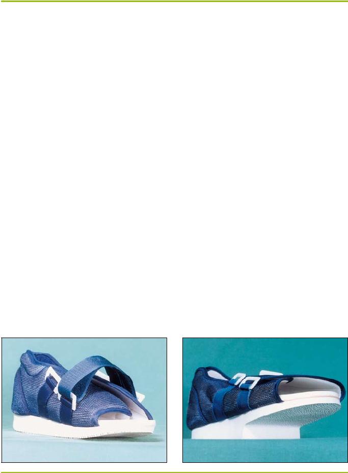
20.Coleman W & Plaia A. Soft moulded sandals for insensitive foot care. Clin Prosthetics & Orthotics 1988; 12(2):67-73.

21.Sarnow M, Eves A, Giurini J, Rosenblum B, Chrzan J & Habershaw G. In-shoe pressure measurements in diabetic patients with at-risk feet and in healthy subjects. Diabetes Care 1991; 17(9):1002-06.

22.Frykberg R, Lavery L, Pham H, Harvey C, Harkless L & Veves A. Role of neuropathy and high foot pressures in diabetic foot ulceration. Diabetes Care 1998; 21(10):1714-19.

23.Novick A, Stone J, Birke J, Brasseaux D, Broussard J, Hoard A & Hawkins E. Reduction of plantar pressure with the rigid relief orthosis. J Amer Podiatric Med Assoc 1993; 83(3):115-22.

24.Landsman A, Meaney D, Cargill R, Macarak E & Thibault L. High strain rate tissue deformation: a theory on the mechanical etiology of diabetic foot ulcerations. J Amer Podiatric Med Assoc 1995; 85(10):519-27.

25.Cochrane G. A Primer of Orthopaedic Biomechanics. New York: Churchill Livingstone, 1982.

26.Brill L, Cavanagh P, Doucette M & Ulbrecht J. Prevention, protection and recurrence reduction of diabetic neuropathic foot ulcers.(number 5 in a series). Treatment of Chronic Wounds (pamphlet).

27.Guzman B, Fisher G, Palladino S & Stavosky W. Pressure-removing strategies in neuropathic ulcer therapy. Cited in Clin Podiatric Med and Surg 1994; 11(2):339-53.

28.Murray H, Young M, Hollis S & Boulton A. The association between callus formation, high pressures and neuropathy in diabetic foot ulceration. Dia- betic Med 1996; 13:979-82.

29.Saye D. The foot: ulcer and toenail debridement and pressure-relief padding. Ostomy/Wound Management 1994; 40(6):14-25.

30.Armstrong D, Liswood P & Todd W. Potential risks of accommodative padding in the treatment of neuropathic ulcerations. Ostomy/Wound Management 1995; 41(7):44-49.

31.Armstrong D & Athanasiou K. The edge effect: how and why wounds grow in size and depth. Clin Podiatric Med & Surg 1998; 15(1):105-08.

32.Chantelau E & Haage P. An audit of cushioned diabetic footwear: relation to patient compliance. Diabetes Med 1994; 11:114-16.

33.Soulier S, Godsey C, Asay E & Perrotta D. The prevention of plantar ulceration in the diabetic foot through the use of running shoes. Diabetes Educator 1987; 13(2):130-32.

34.Nawoczenski D, Birke J & Coleman W. Effect of rocker sole design on plantar forefoot pressures. J Amer Podiatric Med Assoc 1988; 78(9):455-60.

35.Schaff P & Cavanagh P. Shoes for the insensitive foot: the effect of a rocker bottom shoe modification on plantar pressure distribution. Foot and Ankle 1990; 11(3):129-40.

36.Fleischli J, Lavery L, Vela S, Ashry H & Lavery D. Comparison of strategies for reducing pressure at the site of neuropathic ulcers. J Amer Podiatric Med Assoc 1997; 87(10):466-72.

37.Chantelau E, Breuer U, Leisch A, Tanudjaja T & Reuter M. Outpatient treatment of unilateral diabetic foot ulcers with ‘half shoes’. Diabetic Med 1993; 10(11):267-70.

38.Needleman R. Successes and pitfalls in the healing of neuropathic forefoot ulcerations with the IPOS postoperative shoe. Foot and Ankle Inter 1997; 18(7):412-16.

39.Birke J, Novick A, Graham S, Coleman W & Brasseaux D. Methods of treating plantar ulcers. Physical Ther 1991; 71(2):116-22.

40.Petchell A, Keenan A & Landorf K. National clinical guidelines for podiatric foot orthoses. Aust J Podiatric Med 1998; 32(3):97-103.

41.Albert S & Rinoie C. Effect of custom orthotics on plantar pressure distri- bution in the pronated diabetic foot. Foot & Ankle 1994; 33(6):598-604.

42.Brodsky J, Kourosh S, Stills M & Mooney V. Objective evaluation of insert material for diabetic and athletic footwear. Foot and Ankle 1988; 9(3):111-16.

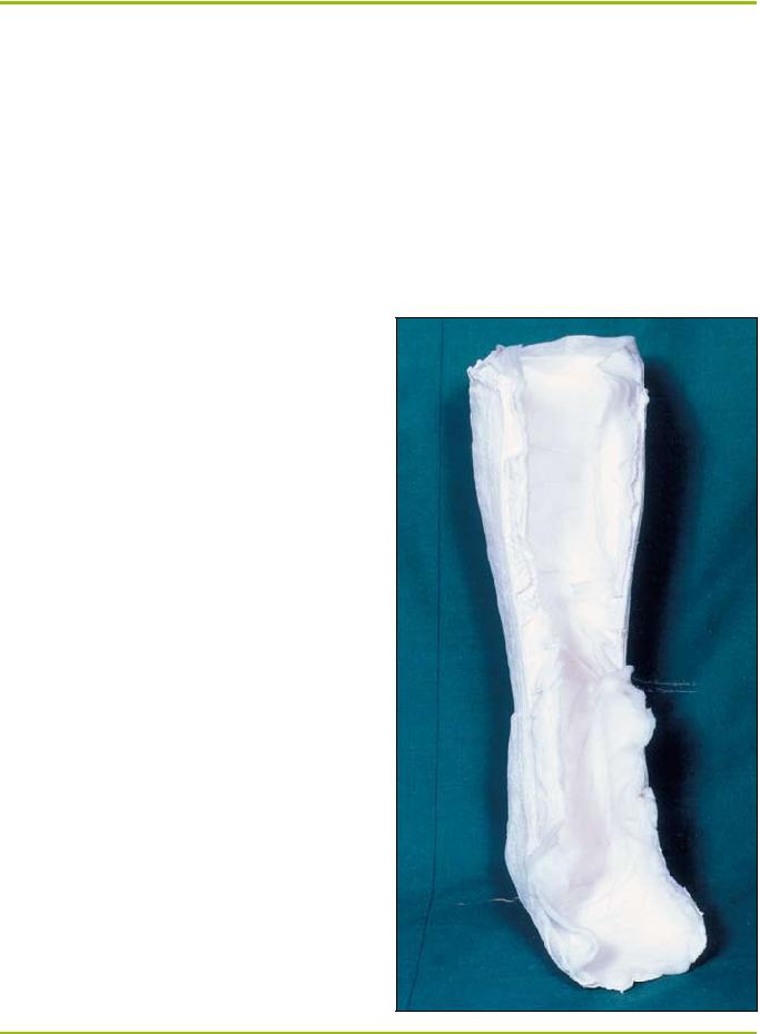
43.Burden C, Jones G, Jones R & Blandford R. Use of the ‘Scotchcast boot’ in treating diabetic foot ulcers. Brit Med J 1983; 28(6):1555-57.

44.Beiser I, Light M, Lynch T & Kanat I. The effect of various forms of immobilisation on plantar foot pressure. J Amer Podiatric Med Assoc 1991; 81(4):206-14.

45.Page S. ‘Neofract’ boots for diabetic neuropathic foot ulcers. J Brit Podiatric Med 1996; 51(2):19-23.

46.Sinacore D, Mueller M, Diamond J, Blair V, Drury D & Rose S. Diabetic plantar ulcers treated by total contact casting. Physical Ther 1987; 67 (10):1543-52.

47.Armstrong D & Lavery L. Evidence-based options for off-loading diabetic wounds. Clin Podiatric Med & Surg 1998; 15(1):95-104.

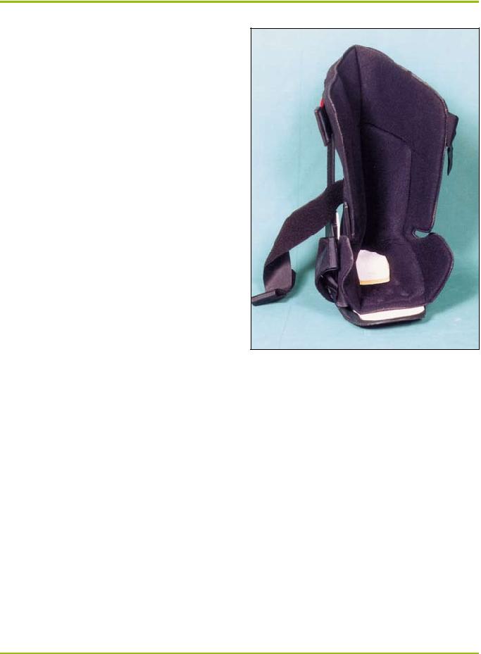
48.Ritz G, Rowland W & Rowland J. Use of the Cam walker in treating diabetic ulcers. J Amer Podiatric Med Assoc 1996; 86(6):253-56.

49.Nawoczenski D, Birke J, Graham S & Koziatek E. The neuropathic foot – a management scheme. Physical Ther 1989; 69(4):287-91.

50.Murray H, Veves A, Young M, Richie D & Boulton A. Role of experimental socks in the care of the high-risk diabetic foot. Diabetes Care 1993; 16 (8):1190-92.

51.Meuller M, Sinacore D, Hoogstrate S & Daly L. Hip and ankle walking strategies: effect on peak plantar pressures and implications for neuropathic ulceration. Arch Phys Med & Rehab 1994; 75(11):1196-1200.

52.Walker S. Sensory substitution in the diabetic neuropathic foot. J Amer Podiatric Med Assoc 1997; 87(7):338.

53.McGill M, Collins P, Bolton T & Yue D. Management of neuropathic

ulceration. J Wound Care 1996; 5(2):52-54.

108 Primary Intention

August 1999